External Invasive Root Resorption Management: A Case Report
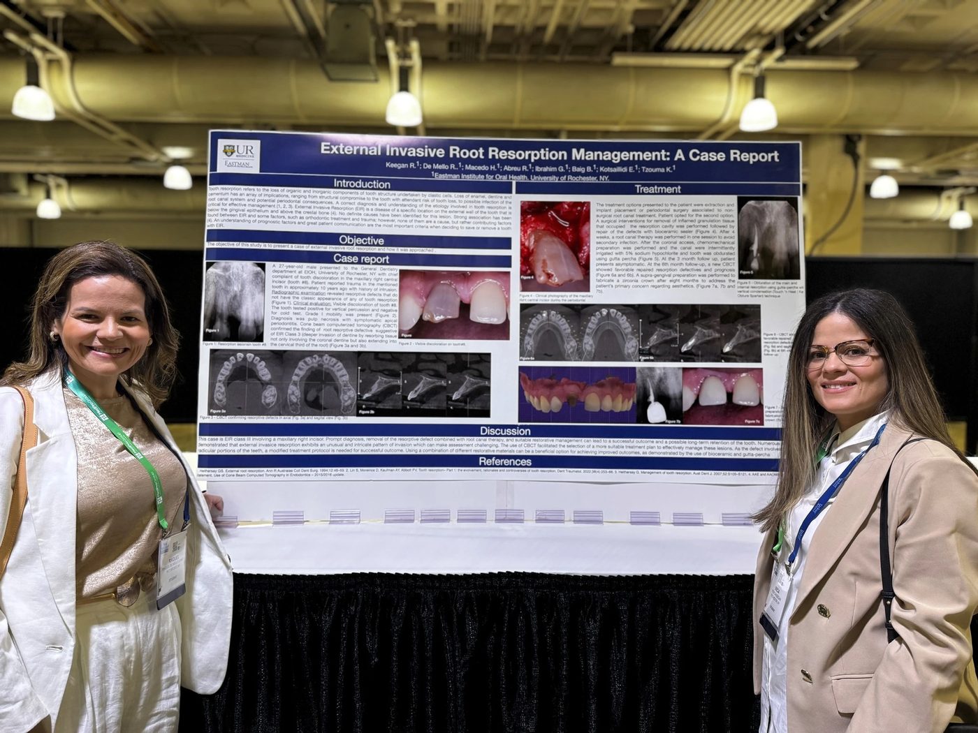
Study title: External Invasive Root Resorption Management: A Case Report Authors: Keegan R, De Mello R, Macedo H, Abreu R, Ibrahim G, Baig B, Kotsaitici E, Tzouma K Institution: Eastman Institute for Oral Health, University of Rochester, NY
AEGD residents Dr. Rebeca Keegan and Dr. Raquel De Mello presented this case at the American Association of Endodontists meeting in Boston (#AAE25).
1. Introduction
External invasive root resorption refers to a progressive loss of cementum and dentin caused by clastic cells. It is considered a rare but aggressive form of external root resorption.
Key characteristics
- Begins on the external root surface
- Often located near the cervical region of the tooth
- May progress into dentin while the pulp can remain initially protected
- Can eventually compromise tooth structure and lead to tooth loss
Etiology and risk factors
The exact cause isn't always identifiable, but associated factors include dental trauma, orthodontic treatment, internal bleaching, periodontal treatment, and surgery or restorative procedures.
Clinical importance
Early diagnosis is critical because once the lesion becomes extensive it may threaten tooth survival.
2. Objective
To present a clinical case of external invasive root resorption and describe the diagnostic process and treatment approach used to preserve the tooth.
3. Case report
Patient: 27-year-old male.
Chief complaint: Tooth discoloration in the maxillary right central incisor (tooth #8).
History: Dental trauma about 10 years earlier; no symptoms at the time of presentation.
Clinical findings: Visible coronal discoloration of tooth #8, sensitivity to percussion, and a negative cold test, findings that suggested pulpal necrosis.
Radiographic examination: A resorptive defect with a lesion extending into dentin, consistent with external invasive root resorption.
Diagnosis: Pulp necrosis with symptomatic apical periodontitis and external invasive root resorption.
Classification: Class 3 external invasive resorption: deep invasion into dentin, extending into the cervical third of the root, requiring more complex treatment.
Imaging: CBCT was used to evaluate the exact size of the lesion, determine the extent of resorption, and plan surgical access.
4. Treatment
Two options were presented to the patient:
- Extraction with implant placement
- Periodontal surgery combined with root-canal treatment
The patient chose tooth preservation with surgical treatment.
Surgical procedure
- Surgical access to expose the resorptive lesion
- Removal of all granulation tissue occupying the resorption cavity
- Restoration of the defect using bioceramic sealer to seal the cavity
Endodontic treatment
After surgical management, root-canal therapy was performed: access-cavity preparation, chemomechanical preparation, irrigation with 5% sodium hypochlorite, and canal obturation using gutta-percha, necessary because the tooth presented with pulp necrosis.
Follow-up
- 3-month follow-up: patient asymptomatic.
- 6-month follow-up: CBCT showed stable repair of the resorption area with no signs of continued resorption.
Final restoration
To address esthetic concerns, a zirconia crown was fabricated, with the restoration completed about eight months after treatment.
5. Results
The treatment achieved successful removal of resorptive tissue, stabilization of the lesion, functional preservation of the tooth, and esthetic restoration with a zirconia crown. The tooth remained asymptomatic with no progression of resorption.
6. Discussion
- Early diagnosis is essential: external invasive root resorption is difficult to detect clinically in early stages.
- CBCT is extremely valuable: three-dimensional imaging allows accurate assessment of lesion size, location, and dentin involvement.
- Combined therapy is effective: combining surgical debridement, root-canal therapy, and modern restorative materials can successfully manage advanced lesions.
- Bioceramic materials improve outcomes: bioceramic sealers provide excellent sealing ability, biocompatibility, and support for healing.
7. Conclusions
- External invasive root resorption can be successfully managed with early diagnosis and appropriate intervention.
- CBCT imaging improves diagnostic accuracy and treatment planning.
- Surgical removal of resorptive tissue combined with endodontic therapy can preserve teeth that might otherwise require extraction.
- Modern materials such as bioceramics and zirconia restorations help achieve lo