CBCT in Endodontics: When 3D Imaging Changes the Diagnosis
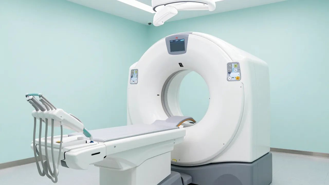
A standard periapical radiograph compresses a three-dimensional tooth into a flat image. Roots overlap, the surrounding bone is superimposed, and subtle disease can hide behind denser structures. Cone-beam computed tomography (CBCT) removes that limitation by reconstructing the area in three dimensions, and in endodontics, that extra information sometimes changes the diagnosis entirely.
What CBCT adds over conventional radiographs
CBCT produces a volumetric data set that can be examined slice by slice in any plane, without the superimposition that limits 2D films. In endodontic practice this is especially useful for:
- Detecting apical periodontitis earlier. Periapical lesions confined within cancellous bone can be invisible on a periapical radiograph until they erode the cortical plate. Studies consistently show CBCT detects these lesions more often than 2D imaging.
- Finding missed canals. Complex anatomy (a second mesiobuccal canal in an upper molar, extra canals, unusual branching) is a leading cause of treatment failure. CBCT helps locate canals that a flat image cannot resolve.
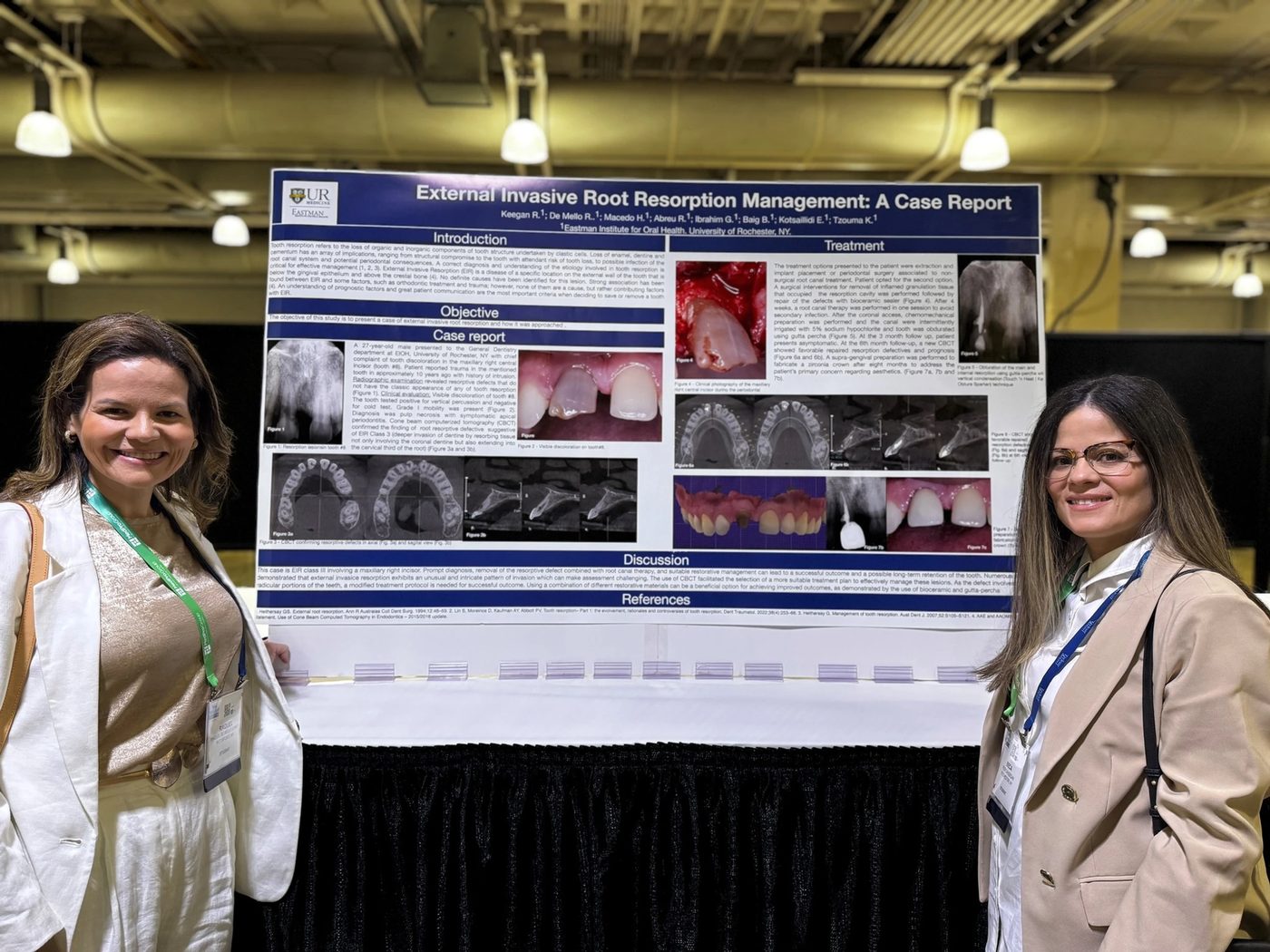
- Assessing resorption. Internal and external (including invasive cervical) resorption can be precisely localized and measured in three dimensions, which directly informs whether and how a tooth can be saved.
- Evaluating trauma and suspected fractures. Horizontal and oblique root fractures, and the relationship of roots to anatomical structures, are far clearer in 3D.
- Planning surgery. Before apical surgery, CBCT clarifies root anatomy and proximity to the sinus or the inferior alveolar nerve.
The guiding principle: selective, not routine
More information is not automatically better. CBCT delivers a higher radiation dose than a periapical radiograph and can be degraded by artifacts. Streaking from metal posts, crowns, and even gutta-percha can obscure exactly the area of interest.
For this reason, the joint position statement of the American Association of Endodontists and the American Academy of Oral and Maxillofacial Radiology frames CBCT as a problem-solving tool, to be used when its expected benefit outweighs the additional dose, not as a routine replacement for conventional imaging. The decision should be case-specific, guided by the principle of using the smallest field of view and lowest dose necessary (the ALARA / ALADA philosophy, as low as reasonably/diagnostically achievable).
In practice, that means CBCT is often justified for situations such as:
- Persistent symptoms when a 2D radiograph is inconclusive
- Suspected extra canals or complex anatomy
- Diagnosis and management of resorption
- Assessment of dental trauma
- Pre-surgical planning and evaluation of treatment outcomes in difficult cases
And it is generally not indicated as a routine screening tool for every root canal.
Limitations to keep in mind
CBCT is powerful but imperfect. Beyond radiation dose and metal artifacts, a limited field of view may miss findings outside the scanned region, and image interpretation requires training. The clinician is responsible for reviewing the entire captured volume, not just the tooth in question. The technology informs clinical judgment; it does not replace it.
The bottom line
Used selectively, CBCT can be the difference between guessing and knowing, revealing a hidden canal, confirming a fracture, or mapping a resorptive defect before deciding whether a tooth can be preserved. The art lies in choosing the right case: enough imaging to answer the clinical question, and no more. That balance of information and restraint is exactly what evidence-based endodontics aims for.
This article is for general educational purposes and is not a substitute for individualized advice from a qualified dentist or endodontist.